Saturday, February 21, 2009

Peizo round tip ?

Dear Dr Huang

This is a problem I have been facing lately; a sloped
sinus floor. For this case I would traditionally drill
to 6mm (about 1mm from the shortest distance of the
floor). But this usually makes osteotome very hard. I
would very nervously take a round bur to go deeper on
the mesial side (but this is very scary).

would peizo machine with a round tip be safer for
these kind of cases

thanks

** Hi Bill:

Nice to see you again !
A case you presented is nice case to practice osteotome sinus lift.
Leave 1 ~2 mm bone (drill preparation to 3.5mm) under sinus cavity plate, then use
medium size osteotome (with some bone graft) tapping into sinus cavity.
After sinus membrane lifting 3~4 mm in length, a little bit shifts mesially to lift sinus membrane
on the slope. More grafts and inserts a implant in.
Please try and hope you get through.
You may take a look my blog about this technic.
http://ajouimplant.blogspot.com/2007/07/osteotome-sinus-lift-augmentation.html
http://ajouimplant.blogspot.com/2007/11/nice-result-for-internal-sinus-lift.html


Good Luck

Dr Perio

Labels: ,

Tuesday, February 12, 2008

Sinus Lift Complication after using Summer’s Ostetome Technique?

Dr. B. asks:
2 months ago I placed an implant in #14 area using a drill to prepare the osteotomy and Summer’s osteotome technique on the apical 3mm to raise the sinus floor. The procedure was uneventful and the area is healing very well. However the patient had emetic episodes on the night of the procedure and has been experiencing vertigo and light headedness since the procedure was performed especially when tilting her head backwards. Anybody have an idea what is going on here? Is any further treatment indicated at this time?

Labels: ,

Friday, January 18, 2008

about Dental implant complications & technological advancements-2

Dr.Mohammad Saeed replied to the questions about Dental implant complications & technological advancements

Greetings Dear Dentist,

A complication like :1: Damaging important anatomical structures such as the sinus or inferior Aleveolar canal could be easily avoided by MRI,which determines the thickness of the canal and the exact position and thickness of the neuro vascular bundle inside.CT also can define it but it fails in 2% of the cases according to C.E Misch.(also determines the thickness of the cortical plate of bone lining the sinus membrane to avoid penetrating it) as well as rare complications such as perforating buccal or lingial plates of bones can be avoided as well .

2: Over heating of bone due to drilling or cooling out side the 'bur-bone contact' or trauma from uncontrolled torque motors ( or controlled but too low RMP) could be avoided by the use of bone expanders ( which i use for more than years now with extreme success even experimentally on dogs)

3:complications of doubting an infected implant can be easily avoided and turned to Absolute sterilization thanks to the Gamma radiation.

4:complications like infection from surrounding roots can be avoided by panoramic and all the more advanced radiological methods.

5:Complication like over bleeding can be avoided thanks to blood sugar tests (which are easly made and fast to help the patient control the sugar level in the blood for about 2 weeks before implant surgeries)

6:complication like overloading can be detected by routine panoramic x rays to observe any resorption may occur in the bone with the direction ( angle ) of overloading.
...wish to continue but i am sorry i have to go.. :) we will continue later inshallah... :)
to know more about my researches please join my group which is facebook dentists...you will find some dogs photos on the main page( researches on implants)..if you have any questions pls let me know..nice to meet you dear Dentist.
bye

about Dental implant complications & technological advancements.

Dr. Anthony G. Sclar repied to the questions about Dental implant complications & technological advancements.

Improvements in implant surface technologies, surgical instrumentation, and prosthetic connections and restorative materials have greatly reduced hardware associated complications. In addition, implant designs which incorporate a “superior shift” of the implant abutment junction such as the Straumann ITI implant or more recently a “central shift” of the implant abutment junction such as the Prevail ™implant from BioMet 3i, the Ankylose® implant from Dentsply Tulsa Dental, and the OsseoSpeed™ implant from Astra Tech Dental, provide the opportunity for improved management of biologic width thus enhancing “soft tissue integration” and stability of underlying crestal bone levels. The potential benefits include improved esthetics and reduced incidence of peri-implant mucositis or peri-implantitis.

Nevertheless, I believe that Cone Beam CT technology and treatment planning software have the greatest potential for helping doctors avoid or reducing the numbers of implant complications. Our in office I-Cat cone beam CT (Imaging Sciences International) scanner allows us to evaluate the patient’s anatomy in 3 D and accurately identify the location and course of vital structures such as the inferior alveolar nerve. In addition, dental pathology not seen on plain films occasionally becomes readily apparent with this technology. When combined with a scan guide derived from a diagnostic wax up that duplicates the proposed final restoration, we are able to perform 3D treatment planning as we evaluate all of the restorative and surgical information on the screen. We can then convert the scan guide into a conventional surgical guide to prepare our sites for implant placement or to guide our 3D hard tissue site development procedures. The 3D diagnostics provides greater information allowing us to make better treatment planning and intra-operative decisions. Taking it a step further, we can order a computer generated surgical guide that incorporates a master cylinder and drill sleeves to allow precise 3D osteotomy preparation or even guided implant placement.

As with any technology, there is a learning curve and nuances which must be understood in order to avoid complications related to the technology itself. Some examples include misinterpretation of CT data or misfit or movement of a guide during surgery which can lead to irreversible complications. The bottom line for doctors is to make a commitment to learn all that they can about the technology and apply it at an entry level before proceeding into advanced applications such as guided surgery.

Thursday, January 03, 2008

Nobel Direct Dental Implants In Question

Three years after patients were given Nobel Direct dental implants, the risk of the implant loosening has increased even more. A follow-up by researchers at the Sahlgrenska Academy in Sweden shows that eight per cent of the implants are lost.
Nobel Direct was launched in 2004 by Nobel Biocare AB. The implants were considered a great innovation, as they could be screwed directly into the jawbone without having to first lift up the mucous membrane.
"We have followed up 48 patients who were among the first to get the implant. For each passing year, we have been able to see how the problems related to these implants have grown more and more," observes Pär-Olov Ostman, a dentist who presented the study at the defence of his dissertation.
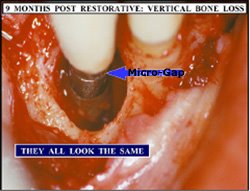
After one year, about five per cent of the implants had been lost, and 20 per cent of the remaining implants showed bone loss of more than three millimetres. The new report indicates that after three years, eight per cent of the implants had been lost, and 25 per cent of the remaining implants showed bone loss of more than three millimetres.
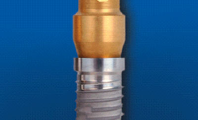
"We believe that the problems related to Nobel Direct result both from the design of the implant and an uneven surface against the soft tissue in combination with the method of treatment recommended by the company," according to Professor Lars Sennerby.
For some time, Nobel Direct was marketed as an implant that was easy to use, and therefore suitable for less experienced dentists. According to the company's marketing, the implant would also counteract marginal bone loss.
"If the implant is inserted in a more conservative manner, avoiding direct load, the results appear to be better. We believe that there are additional implants with similar design that can also cause problems if they are inserted in the same way as Nobel Direct. However, we are unable to draw any certain conclusions regarding these," says Professor Tomas Albrektsson, the head of the Department for Biomaterials Science at the Sahlgrenska Academy.
At the request of the Medical Products Agency, Nobel Biocare AB is now working to clarify certain information in the product information material. The company also markets several other titanium implants that have been scientifically proved to be very safe, including a Brånemark implant with the same patented surface as the Nobel Direct implant in dispute.

----------------------------
Article adapted by Medical News Today from original press release.
----------------------------

Facts about dental implants
The implant is a type of artificial dental root made of titanium. The titanium screw is surgically inserted in the jawbone, and must often become well-secured there so that after several months, it can be used as a foundation for crowns, bridges and dentures. The method was invented by Professor Per-Ingvar Brånemark at the Sahlgrenska Academy in the 1960s. There are several types of titanium appliances, but all are based on titanium being a metal with the unique property of being able to be osseointegrated.
The Sahlgrenska Academy is the health science faculty of Göteborg University. Teaching and research is carried out in medicine, odontology and nursing sciences. We have about 4,000 undergraduate and 1,000 postgraduate students. Of our 1,500 employees, 850 are teachers and/or researchers.
For additional information, please contact:
Elin Lindström Claessen
Information Officer, Sahlgrenska Academy at Göteborg University
Source: Ulrika Lundin
Swedish Research Council

Main Category: Dentistry News
Article Date: 02 Jan 2008 - 0:00 PST

Monday, September 17, 2007

What will you do for this case?

Labels: , ,

Wednesday, August 15, 2007

How to Avoid Hitting the Mental Foramen Nerve?

  1. Dr. Mehdi Jafari Says:

    When we talk about the anterior loop, we mean ‘‘an extension of the inferior alveolar nerve, anterior to the mental foramen’’. Some investigators have also referred to it as the anterior loop of the mental nerve, or described it as the mental neurovascular bundle traversing inferiorly and anteriorly to the mental foramen, which then doubles or loops back to exit the mental foramen. Detection and measurement of the anterior loop was attempted using a variety of diagnostic methods e.g. panoramic films of markers in dried skulls and cadaver mandibles, periapical films of cadaver jaws, and CT scans of patients and surgical cadaver dissections. Clinicians in doubt concerning the position of the mental foramen or who are considering placing an implant in the foraminal region at a depth where there is unease about not having a 2-mm clearance coronal to a location where an anterior loop exists should obtain a CT scan prior to implant placement to avoid injury of the inferior alveolar or mental nerve, however, the mental foramen’s location can be surgically verified. Evidence indicates that an anterior loop is present. It has been detected radiographically and by cadaver dissection; however, its size is debatable. In general, radiographic studies indicated that the anterior loop may be as long as 7.5 mm. Those who are still unwilling to accept the scientific facts can be referred to the following literature:
    1) Solar P et al. A classification of the intraosseous paths of the mental nerve. Int J
    Oral Maxillofac. Implants 1994; 9:339-344
    2) Mardinger O. et al. Anterior loop of the mental canal: An anatomical-radiologic study. Implant Dent 2000; 9:120-125.
    3) Jacobs R et al. Appearance, location, course, and morphology of the mandibular incisive canal: An assessment on spiral CT scan. Dentomaxillofac. Radiol. 2002; 31:322-327.
    4) Kieser J et al. Patterns of emergence of the human mental nerve. Arch Oral Biol 2002; 47:743-747.
    5) Kuzmanovic DV et al. Anterior loop of the mental nerve: A morphological and radiographic study. Clin Oral Implants Res 2003; 14:464-471
    6) Jacobs R, et al. Appearance of the mandibular incisive canal on panoramic radiographs. Surg Radiol. Anat. 2004; 26:329-333.

Wednesday, January 17, 2007

Torn Schneiderian Membrane

Jason, a dental implant patient from New York, asks:

Several months ago I had a Sinus Lift and Bio-Oss was used. The dentist told me that he had torn the Schneiderian Membrane during the procedure. He used 7 grams of Bio-Oss along with Bio-Guide.
I started having very high fevers from the day after the procedure and a major post nasal drip of pus. I was placed on antibiotics and so far I have had 7 rounds of them and still not getting better.
Is it possible that the sinus lift site was overpacked with Bio-Oss. What could happen if the Schneiderian Membrane was not completely fixed? Could that be causing me problems? Should I have the graft material taken out? Any input is greatly appreciated. Thank You

Discussion on OsseoNews:
http://www.osseonews.com/torn-schneiderian-membrane/

Tuesday, November 21, 2006

New Crown Lengthening Techniques and Biologic Width?

A discussion among dental professionals on the message boards of Dentaltown.com.
Log on today to participate in this discussion and thousands more.

tannermck | Total Posts: 3 | Member Since: 08/03/05 | Location: Madison, WI | Posted: 6/19/2006 7:34:42 PM | Post: 1 of 41
Alright, I’ve got questions about perio crown lengthening [CL], now called “gum lifts,” with lasers. To sum it up, if one uses a soft-tissue laser to recontour the free gingival margin, and possibly ablate attached tissue also, what has been done about the osseous crest? The soft tissue may look great immediately, but if
any restorative is going to follow, won’t you be violating the biological width? Setting up multiple long-term problems? If so, it seems as though the soft-tissue laser is really no better than a good electrosurgery unit and not a true replacement for osseous crown lengthening. I am interested in opinions of operators doing various techniques. Periodontists and self-proclaimed cosmetic dentists are invited to weigh in.

Harry J. Jackson | Total Posts: 964 | Member Since: 03/07/05 | Location: Ft. Hood, TX | Posted: 6/19/2006 8:00:32 PM |
Post: 3 of 41
I don’t know who’s calling it “gum lifts,” I hope it’s not doctors! You need to understand the difference between a gingivectomy and crown lengthening. Crown lengthening involves osseous surgery. Some say that they can do this with an envelope-type flap, or even better, a sulcular flap without papilla release. I don’t see how it’s possible! The osseous surgery that is required is so delicate and exacting that I think you need to have x-ray vision to do it well.When CL has been treatment planned it doesn’t matter what you use to make that initial gingivectomy
incision; laser, electrosurge, or my preference, a blade. But, you have to make sure that a flap is reflected and the osseous is done properly. Please, I really hope we don’t start calling anterior esthetic crown lengthening “gum lifts.”

Matt Brink | Total Posts: 3589 | Member Since: 04/28/04 | Location: Wheaton, IL | Posted: 6/20/2006 4:39:59 AM |
Post: 7 of 41
Mike, these are my pictures. Thanks for mistaking them as Danny’s. It made my day. I did the RR [root reshaping] and CL with a bur and photographed the site prior to making bony corrections anywhere else. It shows what you would have to do with a laser. Could it be done closed flap? I sure don’t know how it would be possible.

tannermck | Total Posts: 3 | Member Since: 08/03/05 | Location: Madison, WI | Posted: 6/20/2006 6:02:26 AM |
Post: 12 of 41
Thanks for the comments. Your comments generally agree with my thoughts on the subject. I’d like to
share a few additional thoughts:
1) Many “cosmetic dentists” are using the term “gum lift” routinely now, especially in advertising in my area. I put cosmetic dentists in quotations because every dentist is a cosmetic dentist whether you realize it or not. Indeed, it is a sugar-coated term, but I feel it is misleading.
2) I recommend to all doing any type of osseous surgery using a surgical handpiece, such as an Impact Air. If you do enough using a traditional high-speed, you will have an episode of air emphysema. It’s only a matter of time.

Wednesday, August 09, 2006

Is it possiblly exist "platform switching" in dental implant-abutment

Dental Implant Abutments

The other day, we were looking at a case from another office, and wondering why the abutments were much smaller, or stepped back from the dental implant itself.

This was quite an old case , so we know that this was not intentional. But what was so interesting was that there was no bone loss in this case. There are other recent studies that have shown no bone loss with an abutment that was step backed from the implant itself. What does that tell us? Some manufacturers have already been offering these types of abutments for quite some time. Is this the future direction for abutments? Feel free to add your feedback below.

Discussion on OsseoNews:
http://osseonews.blogs.com/osseodaily/2005/10/dental_implant_.html#c20678445

Platform switching: a new concept in implant dentistry for controlling postrestorative crestal bone levels.

Histologic and radiographic observations suggest that a biologic dimension of hard and soft tissues exists around dental implants and extends apically from the implant-abutment interface. Radiographic evidence of the development of the biologic dimension can be demonstrated by the vertical repositioning of crestal bone and the subsequent soft tissue attachment to the implant that occurs when an implant is uncovered and exposed to the oral environment and matching-diameter restorative components are attached. Historically, two-piece dental implant systems have been restored with prosthetic components that locate the interface between the implant and the attached component element at the outer edge of the implant platform. In 1991, Implant Innovations introduced wide-diameter implants with matching wide-diameter platforms. When introduced, however, matching-diameter prosthetic components were not available, and many of the early 5.0- and 6.0-mm-wide implants received "standard"-diameter (4.1-mm) healing abutments and were restored with "standard"-diameter (4.1-mm) prosthetic components. Long-term radiographic follow-up of these "platform-switched" restored wide-diameter dental implants has demonstrated a smaller than expected vertical change in the crestal bone height around these implants than is typically observed around implants restored conventionally with prosthetic components of matching diameters. This radiographic observation suggests that the resulting postrestorative biologic process resulting in the loss of crestal bone height is altered when the outer edge of the implant-abutment interface is horizontally repositioned inwardly and away from the outer edge of the implant platform. This article introduces the concept of platform switching and provides a foundation for future development of the biologic understanding of the observed radiographic findings and clinical rationale for this technique.

Int J Periodontics Restorative Dent. 2006 Feb;26(1):9-17. Lazzara RJ, Porter SS

Flapless Implant Surgery

Dr. Berg asks:

I am a general dentist placing many of my dental implants. I have recently read about and taken some courses in flapless implant placement using a surgical template.
In addition, I have discussed this technique with some of the other GP’s I know who are placing dental implants, and they have told me that there is very little pain from flapless procedures as compared to laying a full thickness flap. In their view, because there is very little pain involved with the flapless procedures, more patients are accepting dental implants. I was wondering what others thought about this topic. Would flapless implant placement increase the acceptance of dental implant treatment plans with my patients? What are the pros and cons here? Thanks.

More.....See http://osseonews.blogs.com/osseodaily/2006/08/flapless_implan.html

Saturday, June 24, 2006

Is it possible for immediate dental implantation on molar site?

Objective : To evaluate the success rates of immediate and delayed placement of implants with respect to the causes of tooth extraction and implant positions.
Methods : A total of 310 dental implants (immediate implants∶delayed implants=76∶234) were inserted into 80 patients. The types, sizes and positions of the implants and the causes of tooth extraction were recorded. We then investigated the relationship of implant loss with the causes of tooth extraction and placement methods.
Results : A higher failure rate was found for the implants in the posterior region of the maxilla, and when periodontitis was cited as a reason for tooth extraction. The overall success rates were 93.4% and 95.7% in the immediate and delayed implant placement groups, respectively, after a 2-year follow-up. No obvious relationship of success rate was observed with the implant placement method, cause of tooth extaction, and implants’ position.
Conclusion : The immediate placement of implants into fresh extraction sockets could offer advantages over the delayed implant placement. It seems to be a safe and predictable method for patients.
CMJ 2003;116(8):1216-1219

Saturday, April 29, 2006

Superior Esthetics without Micro-leakage of Bacteria and Bone Loss


In the May/June 2006 issue of Implant News & Views, Dr. Donald Callan explains that one of the most perplexing problems of implant placement has been achieving optimal esthetics and that making implants appear as close as possible to natural teeth should be a high priority to accomplish this. Often micro-gaps are created at the implant-abutment junction (IAJ), which resides in close proximity to or just below the bony crest. The presence of the IAJ micro-gap in close approximation to bone is considered to play a major role in peri-implant inflammation and subsequent circumferential bone loss. Leakage of fluids and penetration of the micro-gaps by periodontopathic bacteria leads to the formation of biofilms that are resistant to hygienic efforts or host defense mechanisms.

The PerioSeal?implant [888-446-7126] is designed as a conventional standard dental crown, utilizing a ferrule attachment and dental cement to simplify the restorative phase. This greater surface area contact and better distribution of occlusal force creates a strong connection without a micro-gap to harbor periodontal pathogens. By capturing and sealing the IAJ within the crown, PerioSeal?implants eliminate a potential haven for organisms adjacent to healthy soft tissue. Dr. Callan shows 5 clinical cases to demonstrate this concept. The article is referenced

Thursday, April 06, 2006

Is it necessary to put a resorbable membrane over the Titanium mesh for bone augmentation


Dr. Huang asks:
Is it necessary to put a resorbable membrane over the Titanium mesh + bone grafting for GBR before dental implantation?
Periosteum is a GBR membrane -like tissue for bone augmentation, isn't it?
Are bone graftings with allograft/Xenograft or autogeneous grafts different approaches for this GBR technique? Comments....

Wednesday, March 29, 2006

Dental Implant Impinged On Nerve

Q. Dr. James asks :
I have a patient where dental implants were placed at the lower left first molar and first premolar positions three months following extraction of the posterior teeth. Unfortunately, the anterior dental implant (12mm Straumann RN) slightly impinged the anterior loop of the mental nerve.
Initially the patient reported numbness of the area, but in the days following, developed and electrical current, TENS type pain. Assessment of the site via OPG and CT occurred at 10 and 14 days, and the offending dental implant was removed at 14 days. The discomfort has proven quite debilitating. The patient was placed on Lyrica by his medico, (neuropeptide blocker) and whilst this improved the pain, the side effects are intolerable.
At five month review there has been some resolution in sensory perception, but pain continues. We are currently investigating surgical exploration of the site, and possible pain management with Acupuncture. Any suggestions?

Commend:

I would make a consultation to Dr. Roger A. Meyer from Marietta, Georgia
Posted by: Mar 28, 2006 1:33:55 PM
I'd refer to someone who repairs such cases rather than exploring myself. Reed Day in Phoenix is outstanding as well as Mark Steinberg at Loyola or Ziccardi at Chapel Hill.
Posted by: Dr. Crystal Baxter Mar 28, 2006 1:49:39 PM
The pain should have subsided if the implant was removed after 14 days of placement. There is a possibility of Neuroma formation from injury to the nerve. If the pain is still intense, the patient should have microsurgical nerve repair. Mass General Hospital, Dr. Leonard Kaban has done a lot of these repairs. I have done a few but the patients have long term paresthesia or anesthesia. I do not do these repairs at this time.
Posted by: Gary Wadhwa Mar 28, 2006 2:50:09 PM
I suggest that you contact Dr. Joseph W. Foote at Presbyterian Hopsital, Philadelphia, PA.
Posted by: Mar 28, 2006 4:55:43 PM
I'm oral surgeon and I perform too much surgery about alveolar nerve. My opinion for this case you should wait for 12 months. During this period you can offer B complex vitamins and moisterd hot compress for related area.If alveolar nerve injured, totally nerve reposition or surgery may be needed.
Posted by: Ozyuvaci Hakan Prof.Dr. Mar 28, 2006 10:00:50 PM
In all cases where implant come close to the nerve or impinges it, the first clinical sign is altered sensation from the patient the NEXT day so at once under local Anesthesia often made intraseptally I have always relieve the implant of 1 or 2 turns ( unscrew it) and there is a return to normal after few days and we control and explain to the patientbut the point is we never wait 14 days otherwise the damage is extensive and long lastingI have many cases treated this way and so far we have not lost the implants
Posted by: zeinou Mar 28, 2006 10:46:39 PM

Download from OsseoNews

Friday, February 03, 2006

Is it suitable to osteoporosis pt on bisphosphonates (Fosamax) for dental implantation

Because of the potential for developing osteonecrosis of the jaws, oral surgery procedures are contraindicated in these cases. This means no extractions or dental implants. Going off the drug will not make them better candidates for dental implants because the effects of bisphosphonates lasts for years.

Comments by Larry S.

Let me preface my remarks by saying that this is not legal advice. I recently attended the International Associaation of OMFS meeting in Vienna and there was a symposia on management of the osteonecrosis patient on bisphosphonates. Fortunatly, almost all reported cases are due to the use of IV bisphosphonates (the exact number escapes me but I remember it being less than 50 worldwide on Fosamax and over 1500 reported on the IV drugs). The general consensus of that meeting (again NOT legal advise) was the routine dentoalveolar surgery including implant placement in patients on Fosamax was OK, but contraindicated in those on the IV meds. Obviously those on IV meds have significant medical problems including metastatic disease, lymphoma, ect. As an oral sureon, I don't really have much choice about extracting some teeth on patients on Fosamax, but I do have a "talk" first about risks/benefits.To put it in perspective, Fosamax has been used for years in millions of doses and ther are precious few reports of negative side effects. I think you have to discuss the situation in detail with the patient when you are doing elective surgery.

Wednesday, January 18, 2006

Dental Implant Failure Treament


I have a patient whose dental implants are completely stable but there is Peri-Implantitis. There has to be an eay way to treat this and prevent this type of situation in the future with other patients. The occlusion is fine and their home care is ok. Any thoughts?

Comments>>>

Saturday, January 14, 2006

Foods that may lead to Bad Breath due to volatile sulfur compounds

Food, if it sits out too long will spoil. That action is due to anaerobic bacteria breaking down proteins in that particular food. In milk, the odor of sour milk is caused by relatives of the bugs that create bad breath, when they break down proteins in milk (and all dairy foods). A reaction takes place where 'the bad breath bugs' extract sulfur compounds from the amino acids in these proteins. Specifically, the amino acid Cysteine is converted to Hydrogen Sulfide (the rotten egg smell) and Methionine becomes Methyl Mercaptan (which smells like a cross between old socks and garlic.) More....

Thursday, January 12, 2006

How to control the bleeding before your application of Emdogain?

Dr. Sculean's suggestions from his Taiwan's lecture!

1. Completely remove all infammations (hard/soft tiss);
2. Rinse with normal Saline frequently;
3. Apply Emdogain from apex to crown;
4. Presuture (Mod. Mattress suture);
5. Premixture with bone graft before applied into defects.

Wednesday, January 11, 2006

Dr. Balshi's "The Immediate Loading Protocols" Part three

Osseonews: Dr. Balshi, from your description of your technique it appears that it is critical for you to determine if the implant fixture has achieved adequate primary stability before you deliver the prosthesis.
Dr. Balshi: Achieving primary stability is critical. We need to know that the implant fixture is securely locked into the surrounding bone in the initial stage. Secondary stability will be achieved later over time. If we determine that an implant fixture has not achieved primary stability, we have to treat it differently.
Osseonews: Could you describe your protocol for determining primary stability?
Dr. Balshi: In the majority of current treatment applications insertion torque generally provides the stability quantification. However in research cases we use Resonance Frequency Analysis (RFA). This provides an accurate, quantitative assessment of the quality of the implant fixture to bone interface. In essence we transmit mechanical vibrations to the implant to bone interface. We then monitor changes in resonance frequency. This provides a very accurate assessment of the stability of the implant in the bone.
Osseonews: How do you deploy this system?
Dr. Balshi: We connect a transducer directly to the implant fixture.
Osseonews: In essence you are measuring the stiffness or rigidity of the implant to bone interface.
Dr. Balshi: That is correct.
Osseonews: So with these accurate measurements you can make decisions about primary stability with confidence.
Dr. Balshi: It especially enables us to make decisions about cases where the bone density is less than desirable and where we have to determine whether we should connect the implant fixture to the prosthesis or if we should just allow it more time to integrate more.
Osseonews: This kind of evaluation would be more important for cases with less dense bone.
Dr. Balshi: We would like to all have the perfect bone morphology and density for implant placement. When that is not the case – as it often is – we need to be in a position to derive the most accurate information we can get about the implant to bone interface. This is where the true value of RFA is apparent. This enables us to make the best possible decisions about treatment.
Osseonews: By comparison, tapping on the implant and evaluating the kind of thud it makes to determine primary stability really pales.
Dr. Balshi: Listening for a thud or testing the implant with a torque wrench provides some information. But the RFA approach is non-invasive and provides highly accurate information, which you can quantitatively measure the changes over time, and is an excellent research tool.
Osseonews: For those wanting more information on RFA, where should they look?
Dr. Balshi: The best reference is: A resonance frequency analysis assessment of maxillary and mandibular immediately loaded implants. Balshi SF, et al. International Journal of Maxillofacial Implants 2005;20:584-594. Glenn Wolfinger and I are coa-authors. This provides a description of the technique as well as the science behind it.
Osseonews: It is very clear that computerized technology plays an important role in your protocols.
Dr. Balshi: Yes and it is important for your readers to understand that we do not simply advocate or trying a technique without the proper training. It is essential to understand the techniques in their entirety so you can apply them. We give courses at the Institute in understanding and using the computer technology and software applications. If you do not truly understands how the whole system works, you might want to consider spending more time learning about it before using it.

Dr. Balshi's "The Immediate Loading Protocols" Part two

Interview conducted by Gary J. Kaplowitz, DDS, MA, MEd, ABGD
Comment on this interview by visiting the OsseoNews Blog

OsseoNews (ON): In your protocol you connect the provisional fixed partial complete denture to the abutments that have been permanently torqued down into the implant fixtures. What is your technique for accomplishing this?
Dr. Balshi: After the abutments are torque down, we insert a prosthetic cylinder over the abutment. We then screw down the cylinder with guide pins which project well above the height of the cylinders. We relieve the prosthesis in the area of the abutment and cylinder and cover the abutments with rubber dam fragments to block out any undercuts. We then deliver acrylic around the cylinders and insert the prosthesis. The acrylic locks the cylinder into the acrylic veneer. We unscrew the guide pins and then adjust and polish the surface of the prosthesis. The prosthesis is then ready for delivery.
ON: How do you manage a situation where you place an implant and fail to achieve primary mechanical stability? For example, suppose you have a ‘spinner’ (i.e., an implant that rotates in the osteotomy site).
Dr. Balshi: We have experienced this problem on numerous occasions. We have developed two treatment protocols for spinners. One approach is to place a healing cap and to leave the implant undisturbed until osseointegration is complete. Another approach is to unite the spinners with stable implants that have achieved primary mechanical stability by means of a rigid prosthesis, an all acrylic fixed prosthesis or definitive metal reinforced prosthesis both work well. We have found that after uniting the spinner to the secure and stable framework eventually results in the osseointegration of the spinner. Spinners can be encorporated into an immediate load protocol. We are in the process of publishing an article on the clinical management of spinners.
ON: What about situations where you have grafted the implant site and fail to achieve primary mechanical stability?
Dr. Balshi: Again the critical requirement is to provide a stable and secure prosthesis that locks in the implant. Even with a graft, if the implant is locked into a stable and secure prosthesis, it will undergo osseointegration and can be used in an immediate load protocol.
ON: What are you using for graft material?
Dr. Balshi: We use autogenous bone for our grafts. We harvest bone from the extraction sites or osteotomy sites. We have found that this is the most successful graft material. It is readily available at the time of surgery and easy to harvest and place.
ON: Your orientation appears to favor fixed partial or complete dentures and fixed-detachable partial and complete dentures as opposed to overdentures.
Dr. Balshi: We have found over time, and again after decades of collecting data on our completed cases, that the fixed alternative is to be preferred over the removable. In the long term we have found that overdentures require far more maintenance and are prone to many problems. A screw-retained fixed-detachable partial or complete prosthesis is far more stable.
ON: What about the problem of providing support for the lips and cheek?
Dr. Balshi: Placing the teeth in the proper position is the most critical aspect of achieving proper lip and cheek support. We then fill in area between the teeth and the residual ridge using pink acrylic or porcelain. These prostheses can be designed for great esthetics and hygiene access. This should not represent a contraindication to using a fixed prosthesis

Saturday, December 24, 2005

Can Implant Connect with Natural Tooth?

Doctors who use this philosophy will sometimes connect an implant to a natural tooth. The advantage of this is that by connecting implants to natural teeth, fewer implants are needed to complete the case. This can dramatically reduce the cost of treatment while allowing the patient to have permanent teeth. The disadvantage of this type of treatment is that should a problem arise with either the implant or natural tooth the problem has to be handled differently because the implants and natural teeth are connected. Furthermore, there are limited data regarding the effects of splinting implants to natural teeth. In this regard, it has been reported that intrusion of splinted teeth and pronounced vertical bone loss around implant abutments are potential sequelae;however, the majority of patients, , in one study suffered no adverse effects. Other reports have indicated that connecting implants to teeth in a fixed prosthesis has a good prognosis. A 5-year prospective study designed to compare bridges supported only by implants with bridges supported by both implants and natural teeth within the same patient, noted no higher risk of implant or prosthetic failure for tooth-implant fixed bridges as comparedwith implant-supported bridges.
>>>A case report of natural tooth intrusion when it connected with implant?>>>
>>>Treatment for #33 intrusion:
1. removal of old bridge,
2. #33 orthodontic extrusion,
3. #34 additional dental implant placement,
4. new #33 crown + #34~36 bridge.

Wednesday, December 07, 2005

How to do Tension-free Sutures for Dental Implant or Bone Augmentation?

To obtain tension free primary closure, the buccal full thickness flap periosteum was scored using horizontal releasing incisions (Periosteum releasing) and spreading by Metzenbaum scissors or Scaple after insertion into the tissue. The flap was approximated using Gortex® (Gore, Flagstaff, AZ) sutures or monofilament sutures. These sutures prevent wicking of bacteria into the graft as well as maintain tensile strength. One cannot overly emphasize the importance of establishing and maintaining primary closure following bone-grafting procedures. The sutures were removed two weeks postoperative. The patient agreed to refrain from wearing a prosthesis over this area thus facilitating undisturbed healing.
Tension-free primary sutures technique
(Edentulous Ridge Expansion + Bone Augmentation): Surgery by Perio